How OCT Scans Are Revolutionizing Retina Care
25February2026
The modern field of eye care requires precise diagnostic methods which enable health professionals to identify eye diseases at their earliest stages while they conduct ongoing assessments of all eye diseases present in their patients. The introduction of Optical Coherence Tomography (OCT) has completely changed how doctors assess patients during their eye examinations. The advanced imaging method enables experts to view the internal eye structures with unmatched precision which leads to better patient results through vision preservation.
What Is Optical Coherence Tomography (OCT)?
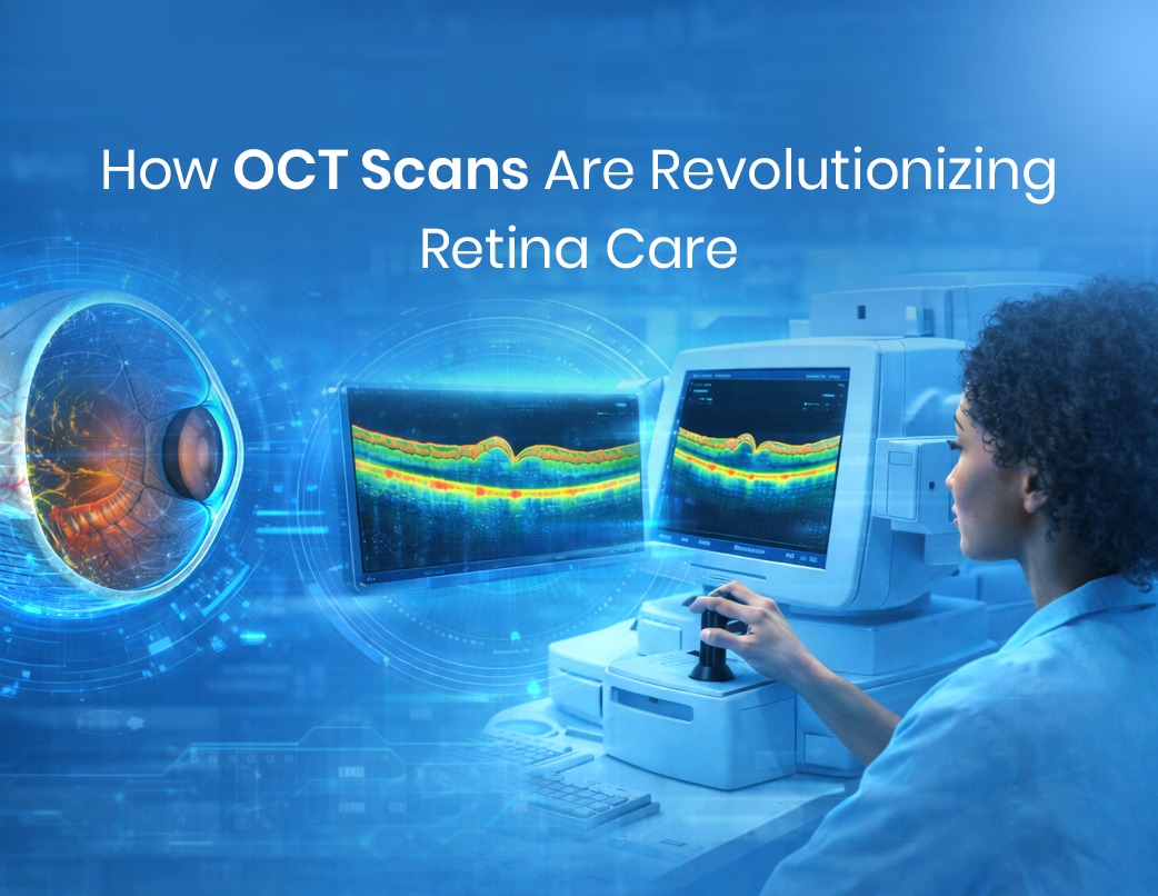
Optical Coherence Tomography is a non-invasive imaging method that uses light waves to create detailed cross-sectional images of the eye. The technology enables ophthalmologists to see the retina and optic nerve plus all essential eye components through their complete structural composition.
OCT imaging enables users to view eye details at a microscopic scale because it records all eye elements without requiring physical contact with the eye which results in a procedure that patients experience as safe, quick and painless.
The current technological development of Swept-Source OCT and OCT Angiography (OCTA) results in improved blood flow visualisation through the eye while providing deeper eye tissue penetration and rapid scanning capabilities.
Why OCT Is Essential in Eye Care
OCT has become a routine and indispensable part of modern ophthalmology because it enables early diagnosis and precise disease management. The majority of eye conditions progress in silence until they reach the point where vision deterioration becomes evident to patients. The OCT system discovers these modifications which occur before the patient reaches the stage of vision loss.
Role of OCT in Retinal Disorders
Retinal diseases represent one of the main causes which lead to worldwide vision loss. The technology of OCT systems plays a crucial role in the detection and treatment of these medical conditions.
- The OCT system enables doctors to observe initial macular changes and fluid buildup which occurs during age-related macular degeneration (AMD) so that they can start treatment at the right moment.
- Diabetic retinopathy treatment requires OCT technology, which helps doctors track retinal swelling, and damage assessment from diabetes, to commence early treatment.
- OCT provides clear imaging of retinal fluid accumulation in macular oedema, which enables ophthalmologists to determine disease intensity and patient response to therapy.
- OCT technology functions as an essential tool for glaucoma detection and patient monitoring throughout their treatment. Glaucoma progressively damages the optic nerve which remains undetected until it reaches later stages.
- The technology of OCT allows doctors to determine optic nerve damage through retinal nerve fibre layer (RNFL) and ganglion cell complex measurements.
- OCT serves as the standard tool for corneal imaging which doctors use to examine corneas before and after LASIK and corneal transplant procedures.
- OCT technology delivers precise measurements for corneal thickness and curvature, and structural corneal integrity assessment.
- OCT technology serves as a crucial tool for retinal surgical procedures which include vitrectomy and retinal detachment repair.
- OCT delivers essential information to doctors who perform retinal surgery, as it helps them assess patients before and after their procedures.
OCT Angiography: Seeing Blood Flow Without Injections
The OCT Angiography system develops through its OCT technology foundation which enables researchers to create high-resolution retinal and choroidal blood flow images without using intravenous contrast material. The procedure guarantees patients experience safer and more comfortable treatment results. OCTA provides effective diagnostic support for eye diseases associated with blood vessel problems which include diabetic retinopathy and retinal vein occlusion and other blood circulation disorders that affect the eye.
The Future of OCT in Ophthalmology
The development of optical coherence tomography continues to progress through artificial intelligence-powered imaging systems and faster scanning technologies and new portable scanning equipment. The new technologies will provide easier access to optical coherence tomography because they will operate at higher efficiency levels while maintaining precise results across various geographical locations.
OCT technology will become more important in eye disease detection because it will enable doctors to develop unique treatment approaches according to each patient’s needs.
Conclusion
OCT is widely regarded as the gold standard in retinal imaging. As it has transformed ophthalmology through its ability to provide safe, accurate and complete internal eye structure visualisation. The combined capabilities of OCT for retinal disease detection, glaucoma assessment, surgical guidance and post-surgical monitoring create a vital resource for contemporary eye healthcare. The system enables early detection of visual changes which allows tracking of disease development to safeguard eyesight while enhancing patient quality of life.
The study of OCT through ophthalmology courses and clinical training programs offers essential knowledge for students and professionals who aim to become experts in advanced diagnostic methods.
Read more here :
Retinal Diseases: Early Detection Can Save Your Vision